Tag Archives: digestive diseases

Swallowing and Dysphagia (with Animation)

Swallowing

Below is a narrated animation about swallowing reflex, phases and overview of neural control. Click here to license this video on Alila Medical Media website.

Swallowing, or deglutition, is the process by which food passes from the mouth, through the pharynx and into the esophagus. As simple as it might seem to healthy people, swallowing is actually a very complex action that requires an extremely precise coordination with breathing since both of these processes share the same entrance: the pharynx. Failure to coordinate would result in choking or pulmonary aspiration. Swallowing involves over twenty muscles of the mouth, throat and esophagus that are controlled by several cortical areas and by the swallowing centers in the brainstem. The brain communicates with the muscles through several cranial nerves.

Swallowing consists of three phases

1. Oral or buccal phase: this is the voluntary part of swallowing, the food is moistened with saliva and chewed, food bolus is formed and the tongue  pushes it to the back of the throat (pharynx). This process is under neural control of several areas of cerebral cortex including the motor cortex.
2. Pharyngeal phase starts with stimulation of tactile receptors in the oropharynx by the food bolus. The swallow reflex is initiated and is under involuntary neuromuscular control. The following actions are taken to ensure the passage of food or drink into the esophagus:
– The tongue blocks the oral cavity to prevent going back to the mouth.
– The soft palate blocks entry to the nasal cavity.
– The vocal folds close to protect the airway to the lungs.The larynx is pulled up with the epiglottis flipping over covering the entry to the trachea (the windpipe). This is the most important step since entry of food or drink into the lungs may potentially be life threatening.
– The upper esophageal sphincter (UES) opens to allow passage to the esophagus.
3. Esophageal phasefood bolus is propelled down the esophagus by peristalsis – a wave of muscular contraction that pushes the bolus ahead of it. The larynx moves down back to original position.

Click here to see an animation of the swallowing process on on Alila Medical Media website where the video is also available for licensing.
Swallowing, labeled
Fig. 1: Anatomy of swallowing. See text for details of phases. The blue arrows represent breathed air. Click on image to see a larger version on Alila Medical Media website where the image is also available for licensing.


Dysphagia (swallowing disorders)


This  video is available for licensing on Alila Medical Media website. Click HERE!

Dysphagia refers to a group of conditions characterized by difficulty swallowing. There are two main classes of problems that can lead to swallowing disorders:


 1. 
Neuromuscular problems: 

– Muscular disorders that affect skeletal muscles, such as muscular dystrophy, myasthenia gravis…

– Diseases of the nervous system that compromise the way the brain controls the swallowing reflex, such as stroke, Parkinson’s disease, multiple sclerosis…

Weakened muscles and/or impaired coordination as a result of aging.

This class commonly affects the first two phases of swallowing.

2. Narrowing of the throat or esophagus due to throat cancer, esophageal cancer and formation of small sacs or rings in the walls of the esophagus.  Gastroesophageal reflux disease – GERD – is also a common cause. In GERD, scars resulted from stomach acid injuries may obstruct the esophagus and cause difficulty swallowing.

This class mostly affects the third phase of swallowing.

Schatzki ring blocks esophagus
Fig. 2: Schatzki ring makes the lumen of esophagus smaller. Click on image to see a larger version on Alila Medical Media website where the image is also available for licensing.


For people with dysphagia, eating becomes a challenge. The consequences may be serious. Someone who cannot swallow safely is at high risks of choking, pulmonary aspiration and may not be able to eat enough to stay healthy.

Treatment depends on the cause of the condition:

– Muscle strength and coordination exercises may be recommended for some.

– A change in the position of the head and neck when eating could be beneficial to others.

– Right choice of food and drink is important for most. Soft textured food and thickened drinks are recommended for safe swallowing.

– Surgery may be needed to remove narrowed parts of the esophagus.

– Finally, patients with severe dysphagia and recurrent aspiration may have to resort to tube feeding to get nutrition to the body.

                                                                                                                            See all ENT topics

                                                                                                                    See all Digestive topics

Email this to someoneShare on FacebookTweet about this on TwitterShare on Google+Share on LinkedIn

Vertical Sleeve Gastrectomy and Gastric Lap Band Surgeries

This is a continuation to the main article about bariatric surgery .

Below is a narrated animation of Gastric Sleeve and Gastric Lap Band procedures. Click here to license this video on Alila Medical Media website.

Vertical sleeve gastrectomy (VSG)

In this procedure a cut is made vertically and the larger part of the stomach (up to 85% of its volume) is removed from the body. The remaining is closed with staples to create a “new stomach” that is now having the shape of a tube (see Fig. 1). The procedure preserves both sphincters at the two ends of the stomach and therefore has minimum effect on the functioning of the digestive process.This reduces the risk of malabsorptive complications commonly associated with intestinal bypass. The procedure is irreversible.
Click here to see an animation of VSG procedure  on Alila Medical Media website where the video is also available for licensing.

Vertical Sleeve Gastrectomy (VSG)
Fig. 1: Vertical sleeve gastrectomy procedure. Click on image to see a larger version on  Alila Medical Media website where the image is also available for licensing.


VSG procedure is gaining more and more popularity thanks to its simplicity and good results on initial weight loss. However, due to the lack of long-term data, it is yet to be endorsed by bariatric surgery societies and is not covered by some insurance companies.


Adjustable gastric band (Lap band)


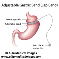
In this procedure an inflatable silicon band is placed around the top of the stomach to create a small stomach pouch (Fig. 2). During a meal, the pouch is filled up quickly with a small amount of food and releases it slowly into the lower part of the stomach due to the restriction by the band. As the pouch is full, it gives a feeling of satiety (fullness). Slow passage of the food makes the patient feel full for a longer period of time and thus reduces the amount of food intake. The band is connected to a port placed under skin of the abdomen. Through this port, a saline solution (salt water) is injected to adjust the diameter of the band and thus making the passage between the pouch and the lower part of the stomach smaller or larger accordingly to the needs of patient.

Click here to see an animation of gastric band procedure  on Alila Medical Media website where the video is also available for licensing.

Gastric Band Weight Loss Surgery


Fig. 2: Adjustable lap band procedure. Click on image to see a larger version on Alila Medical Media website where the image is also available for licensing.


As the procedure involves no cutting or stapling of the stomach, it is minimal invasive and reversible. The surgery can be performed laparoscopically (as opposed to open surgery) through small incisions with the aid of a camera. Recovery time is significantly shortened compared to gastric bypass procedures. Also, as there is no intestinal bypass, the risks of nutritional deficiencies, dumping syndrome and other complications associated with it are significantly reduced.
In terms of weight loss efficiency, however, patients who undergo gastric lap band surgery typically lose less weight than those who have had gastric bypass procedures.

                                                                                              > See Gastric bypass procedures

                                                                                                           > See all Digestive topics

Email this to someoneShare on FacebookTweet about this on TwitterShare on Google+Share on LinkedIn

Bariatric surgery

Below is a narrated animation of body mass index and Roux-en-Y gastric bypass. Click here to license this video on Alila Medical Media website.

Bariatric surgery, or weight loss surgery, refers to a variety of surgical procedures for treatment of morbid obesity. Obesity is determined by Body Mass Index (BMI) which is calculated as the ratio of body weight over square of body height. The higher the BMI the higher the extend of obesity.  A normal BMI is between 20 and 25. An individual is considered morbidly obese if he or she has a body mass index of 40 or more, or of 35 or more and with obesity-related health problems such as diabetes, sleep apnea or hypertension.
Weight loss is achieved by reducing the size of the stomach. Smaller stomach makes you feel full faster and therefore makes it easier to reduce the amount of food intake.

Roux en-Y Gastric bypass (RNY)

This is the most commonly performed bariatric surgical procedure and is considered the gold standard for weight loss treatment. This procedure includes two steps:
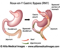
1. The stomach is divided into two part : one small pouch at the top of the stomach where it is connected to the esophagus (gastric pouch in Fig. 1) and the rest of the stomach which will be “bypassed”, the two parts are separated and stapled.
2. Rerouting of the intestine: the intestine is cut at about 45cm (18in) down from the end of the stomach. The first part of the intestine (the duodenum) will be “bypassed”. The top end of the second part (the jejunum) is pulled up and connected to the gastric pouch created in step 1. The lower end of the duodenum is reconnected to the jejunum at a lower point (Fig.1). The new configuration has a shape of an Y, hence the name of the procedure.

Click here to see a video animation of gastric bypass procedure on Alila Medical Media website where the video is also available for licensing.

Roux-en-Y Gastric Bypass (RNY) surgery
Fig. 1: Roux-en-Y gastric bypass diagram. Note the passage of food and digestive juice after surgery. Click on image to see a larger version on Alila Medical Media website where the image is also available for licensing.


How weight loss is achieved?
Firstly, the volume of the stomach is now greatly reduced to a small pouch (usually less than 10% of the original volume) which is filled up fast after a small amount of food intake. This sends a signal to the brain that the stomach is full and generates a feeling of fullness (satiety). This helps to stop eating. Also, if eating continues, vomiting and discomfort may follow. Secondly, as the first part of the intestine (the duodenum) is bypassed, the amount of nutrition absorbed by the body is greatly reduced. In normal digestion, this is where most of the nutrition is absorbed. Malabsorption contributes to weight loss effect.

Complications
While this procedure is proven as an efficient long term weight loss treatment, it associates with significant complication risks. These include: leakage along the staple lines and surgical connections leading to infection and abscess formation; stricture and obstruction of digestive tract due to scar formation; dumping syndrome; nutritional deficiencies; and other general surgical risks  due to complexity of the procedure.

Mini Gastric bypass (MGB)

This is a modification of the more common RNY procedure described above. Here are the differences: (see Fig. 2)
1. In step 1 a long tube is created instead of a pouch.
2. In step 2, the intestine is NOT cut, it is pulled up and hooked up with the new stomach tube.

Mini gastric bypass surgery

Fig. 2: Mini gastric bypass diagram. Click on image to see a larger version on Alila Medical Media website where the image is also available for licensing.


Click here to see a video animation of mini gastric bypass procedure on Alila Medical Media website where the video is also available for licensing.

This procedure is becoming more and more popular as it produces good weight loss results and is simpler than the original procedure resulting in less complication risks. Less cutting and stapling lowers the risk of leakage and infection. It also reduces the bile reflux possibility  as the intestinal rerouting is set at a lower point on the stomach.

                                                                         > NEXT: Gastric Sleeve and Gastric Lap Band

                                                                                                            > See all Digestive topics


Email this to someoneShare on FacebookTweet about this on TwitterShare on Google+Share on LinkedIn

GERD and Heartburn (with video)

Gastroesophageal reflux disease

This video and other animations of the digestive system are available for licensing on Alila Medical Media website. Click here!


Gastroesophageal reflux disease (GERD) or gastric reflux disease is  a chronic condition where acid from the stomach flows up and damages the mucosa of the esophagus.
At the junction between the esophagus and the stomach is the lower esophageal sphincter (LES). The LES is a ring of muscle that is generally closed tight to prevent stomach acid from coming up. In normal digestion, the LES opens shortly to allow food bolus passing down to the stomach and closes back tight instantly. GERD occurs when the LES is abnormally relaxed and can not close properly (Fig. 1). Heartburn is a burning sensation in the chest associated with each regurgitation of gastric acid and is the most prominent symptom of GERD. 

Gastric reflux, labeled diagram.
Fig. 1: Abnormal relaxation of the lower esophageal sphincter as cause of GERD. Click on image to see a larger version on  Alila Medical Media website where the image is also available for licensing.


Hiatus hernia is believed to be another cause of GERD. Hiatus hernia or hiatal hernia is a condition where the top portion of the stomach is pulled up forming a herniation above  the diaphragm. This situation somehow compromises the esophagus – stomach barrier and facilitates acid reflux.

Hiatal Hernia
Fig. 2: Types of hiatal hernia. Click on image to see a larger version on  Alila Medical Media website where the image is also available for licensing.


Treatment

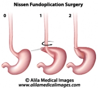
Treatment includes dieting and medication. Proton-pump inhibitors, which act to reduce gastric acid production, are drugs of choice. If these fail, a surgery may be recommended. In a procedure called Nissen fundoplication, the top portion of the stomach is wrapped around the lower part of esophagus and sewn into place. This way, the muscles in the wall of the stomach reinforce the closure of the esophagus. This surgical procedure is particularly recommended when hiatus hernia is present as this can be fixed at the same time. The procedure can be done with minimal invasive laparoscopic technique through small incisions with the aid of a camera.
Nissen Fundoplication Surgery
Fig. 3: Nissen fundoplication procedure.  Click on image to see a larger version on Alila Medical Media website where the image is also available for licensing.


                                                                                                                 > See all Digestive topics

Email this to someoneShare on FacebookTweet about this on TwitterShare on Google+Share on LinkedIn

Digestive System Gallery

Download diagrams (labeled and non-labeled)of organs of human gastrointestinal tract, normal anatomy and diseases, conditions, surgery treatment, and more.

Please note: Free downloads are intended to facilitate healthcare education for people in need in low income countries and can be used for educational purposes only. If you can afford it or if you plan to use the images for commercial purposes, please consider buying instead. You can find a larger image collection at higher resolutions for sale at affordable prices on Alila Medical Media website.

To download: right click on full size image – choose “Save image as” and save it into your computer. By downloading from this website you acknowledge that you agree to our Conditions of Use.
To purchase larger sizes of the image: (left) click on full size image.

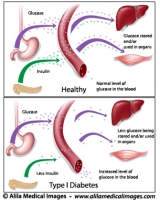
Type 1 diabetes, labeled diagram.
Type 1 diabetes, insulin dependent: pancreas does not produce enough insulin, medical drawing.
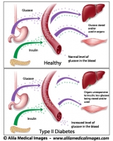
Type 2 diabetes, labeled diagram.
Type 2 diabetes, insulin resistance, pathology of the disease, labeled drawing.
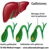
Gallstones, labeled diagram.
Gallstones blocking bile duct, a digestive condition, medical illustration.
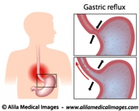
Heartburn, stomach acid reflux, , medical illustration.
Relaxed lower esophageal sphincter (LES) causes gastric reflux and heartburn sensation, medical drawing unlabeled.
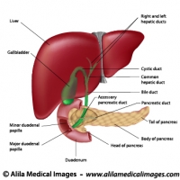
Digestive organs and bile ducts, labeled diagram.
Diagram of human liver, gallbladder, duodenum and pancreas anatomy, labeled.
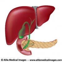
Digestive organs, medical drawing.
Diagram showing anatomy of liver pancreas gallbladder and bile ducts, unlabeled version.
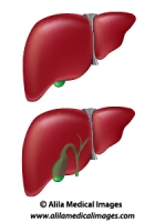
Liver and gallbladder, medical illustration.
Human liver and gallbladder normal anatomy diagrams, two different views, unlabeled.
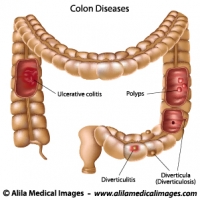
Diseases of the large intestine, labeled diagram.
Illustration of colon pathologies including polyps, colitis, diverticulitis, diverticula, labeled diagram.
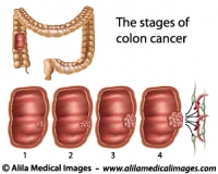
Stages of colon cancer, medical drawing.
Cancer of the large intestine staging diagram, unlabeled.
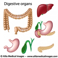
Digestive organs isolated, medical drawing.
Liver, stomach, pancreas, gallbladder, intestine anatomy, unlabeled.
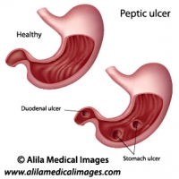
Peptic ulcers, labeled diagram.
Anatomy of healthy stomach and that with peptic ulcers : stomach ulcer and duodenal ulcer.
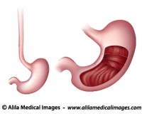
Gross anatomy of stomach, medical drawing.
Diagram of gross anatomy of human stomach showing three layers of muscles in the stomach wall and gastric rugae, unlabeled.
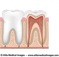
Tooth anatomy, unlabeled diagram.
Tooth anatomy from outside and in cross section, unlabeled drawing.
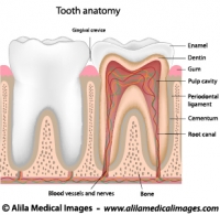
Tooth anatomy, labeled diagram.
Tooth anatomy from outside and in cross section, labeled drawing.
Dental checkup icon
Dental checkup icon
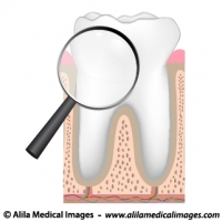
Magnifying glass on a molar tooth, illustration.
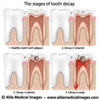
Plaque and tooth decay, medical drawing.
Tooth decay stages diagram.
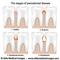
Periodontal Disease, labeled diagram.
Gum disease stages, from gingivitis to periodontitis.
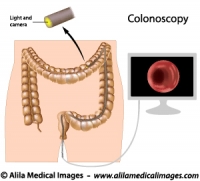
Colonoscopy procedure, labeled diagram.
Endoscopy procedure for diagnosis of colon diseases.
Nissen Fundoplication Surgery, medical drawing.
Nissen fundoplication surgical procedure for treatment of heartburn and hiatus hernia, illustration, unlabeled.
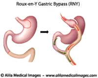
Roux-en-Y Gastric Bypass, medical drawing.
Roux-en-Y Gastric Bypass (RNY) surgery diagram, unlabeled version.
Roux-en-Y Gastric Bypass surgery, labeled diagram.
Illustration of Roux-en-Y Gastric Bypass (RNY) surgical procedure, labeled.
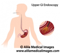
Upper GI endoscopy, medical illustration.
Upper gastrointestinal tract endoscopy for diagnosis of esophagus and stomach disorders, unlabeled.
Gastric Band Weight Loss Surgery, labeled diagram.
Adjustable lap band surgical procedure illustration, labeled drawing.
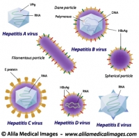
Hepatitis viruses comparison, labeled diagram.
Structure of hepatitis A, B, C, D and E viruses, medical illustration.
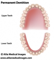
Adult dentition, medical drawing.
Permanent teeth, adult dentition, upper and lower jaws, unlabeled.
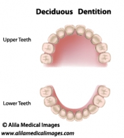
Deciduous dentition, medical drawing.
Deciduous dentition (baby or milk teeth), upper and lower jaws, medical illustration unlabeled..
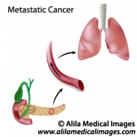
Metastatic cancer diagram
Cancer spreads through blood circulation from pancreas to lungs, unlabeled.
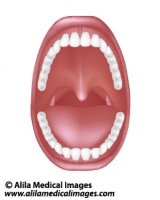
Mouth and throat anatomy, medical drawing.
Diagram of human oral cavity with denture, tonsils, unlabeled.


Email this to someoneShare on FacebookTweet about this on TwitterShare on Google+Share on LinkedIn