Tag Archives: skin problems

Cellulite

The following video is available for licensing on Alila Medical Media website. Click here!


What is cellulite?

Cellulite is the dimpling, lumpy appearance of the skin, commonly occurs in females after puberty age. It’s most visible on the thighs, the buttocks, and belly. Other names include  adiposis edematosa, dermopanniculosis deformans, status protrusus cutis, gynoid lipodystrophy, orange peel syndrome and cottage cheese skin. Cellulite is not a disease and should NOT be confused with cellulitis, which is the infection of skin and underlying tissues.

Anatomy of cellulite

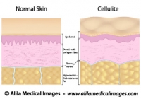
The skin has three layers : epidermis (outermost), dermis and subcutaneous fat (Fig.1). Vertical bands of connective tissue called fibrous septae (singular: septum) connect the dermis to underlying soft tissues. Cellulite happens when fat cells accumulated in the subcutaneous fat layer push the skin up while the fibrous septae pull it down. These two actions in opposite directions result in the bumpy appearance of the skin. In people with thin skin, this becomes even more noticeable.
Cellulite versus smooth skin
Fig. 1 : Structure of normal skin and skin with cellulite, back to back for comparison. Note the fibrous septae pull the skin down in cellulite. Click on image to see a larger version on Alila Medical Media website where the image is also available for licensing.


Causes

Causes of cellulite are not fully understood but the following factors are likely to be involved:
Hormonal : Over 80% of women over the age of 20 has some degree of cellulite. Cellulite is rare in men, but is more common in those with androgen deficiency.
Genetic: Some genetic make-ups are likely to be predisposing factors. You have more chance of getting it if other women in the family have it.
Lifestyle: Diet and exercises definitely have a good share of contribution. Reducing body fat typically improves cellulite appearance.  Extreme diet, however, may produce adverse effect as thinner skin makes it more visible (see the anatomy part above).

Treatment

Various therapies are available including massages, heat therapy, ultrasound, drugs,… These treatments supposedly act to either reduce subcutaneous fat or thicken the skin, but none are scientifically proven to be effective in the long term.

The latest technology based on releasing of the fibrous septae that pull the skin down (see the anatomy section above) has received a better response from scientists. Cellulaze, a device that uses laser beams to cut through the fibrous septae, has produced promising initial results in U.S. clinical trials. It’s been advised, however, to take this approach with precaution given the newness of the technique and shortness of long term data.

Finally, as repetitive and obvious as it may sound, the best treatment for cellulite is to maintain a healthy lifestyle, eating healthy (but no extreme diet), drinking lots of fluid and daily exercises.

                                                                                                  >See all dermatology topics

Email this to someoneShare on FacebookTweet about this on TwitterShare on Google+Share on LinkedIn

Laser skin resurfacing

What is  laser skin resurfacing?

Laser skin resurfacing, also known as laser peel or laser lasabrasion, is a procedure using laser beams to reduce certain skin imperfections such as wrinkles, pigment spots, scars and blemishes.

Below is a narrated animation about laser skin resurfacing procedures. Click here to license this video and/or other dermatology related videos/images on Alila Medical Media website.

How does it works?

The skin is composed of three layers (Fig.1, left panel): epidermis (the outermost), dermis and hypodermis (subcutaneous fat). The dermis contains bundles of well organised collagen fibers which contribute to the firmness and smoothness of the skin. As skin gets older, these fibers become less in number and also less organized, wrinkles and age spots (uneven pigmentation) appear (right panel of Fig. 1).

Wrinkled skin versus smooth skin
Fig. 1 : Structure of young skin and older skin back to back for comparison. Note the differences in collagen fibers number and arrangement. Click on image to see a larger version on Alila Medical Media website where the image is also available for licensing.


Laser beams ablate (destroy) the outer layer of the skin removing unwanted wrinkles and spots. At the same time, the heat of the beams stimulates the growth of collagen fibers in the dermis. As the wound is healing, new skin that grows over it is smoother and firmer (Fig. 2)

Classic laser skin resurfacing versus Fractional laser skin resurfacing

The classic laser skin resurfacing uses laser to ablate a large area of the “problematic” skin, the whole problem (e.g. a dark spot) is removed, the skin is the left to heal naturally by itself. The plus : as the whole “problem” is removed, only one treatment is needed. The minus: burned-out area is large, it takes a long time to heal and is subject to higher risk of infection.
Click here to see an animation of  laser skin resurfacing procedure on Alila Medical Media website where the video is also available for licensing.
Laser Skin Resurfacing, labeled diagram.

Fig. 2 : Laser skin resurfacing technique for removal of a dark spot. Note a large wound after treatment. Click on image to see a larger version on Alila Medical Media website where the image is also available for licensing.


The newer technique : fractional laser skin resurfacing uses multiple smaller laser beams to ablate the skin in smaller spots, leaving undamaged skin tissue in between. The plus: healing is faster and less complicated. The minus: a series of treatments is needed to eliminate the “problem”. See the animation of this procedure here
Fractional Laser Skin Resurfacing
Fig. 3 : Fractional laser skin resurfacing technique. Note smaller wounds after treatment but part of the dark spot still remains after healing.
Click on image to see a larger version on Alila Medical Media website where the image is also available for licensing.

                                                                                                       >See all dermatology topics

Email this to someoneShare on FacebookTweet about this on TwitterShare on Google+Share on LinkedIn

GERD and Heartburn (with video)

Gastroesophageal reflux disease

This video and other animations of the digestive system are available for licensing on Alila Medical Media website. Click here!


Gastroesophageal reflux disease (GERD) or gastric reflux disease is  a chronic condition where acid from the stomach flows up and damages the mucosa of the esophagus.
At the junction between the esophagus and the stomach is the lower esophageal sphincter (LES). The LES is a ring of muscle that is generally closed tight to prevent stomach acid from coming up. In normal digestion, the LES opens shortly to allow food bolus passing down to the stomach and closes back tight instantly. GERD occurs when the LES is abnormally relaxed and can not close properly (Fig. 1). Heartburn is a burning sensation in the chest associated with each regurgitation of gastric acid and is the most prominent symptom of GERD. 

Gastric reflux, labeled diagram.
Fig. 1: Abnormal relaxation of the lower esophageal sphincter as cause of GERD. Click on image to see a larger version on  Alila Medical Media website where the image is also available for licensing.


Hiatus hernia is believed to be another cause of GERD. Hiatus hernia or hiatal hernia is a condition where the top portion of the stomach is pulled up forming a herniation above  the diaphragm. This situation somehow compromises the esophagus – stomach barrier and facilitates acid reflux.

Hiatal Hernia
Fig. 2: Types of hiatal hernia. Click on image to see a larger version on  Alila Medical Media website where the image is also available for licensing.


Treatment

Treatment includes dieting and medication. Proton-pump inhibitors, which act to reduce gastric acid production, are drugs of choice. If these fail, a surgery may be recommended. In a procedure called Nissen fundoplication, the top portion of the stomach is wrapped around the lower part of esophagus and sewn into place. This way, the muscles in the wall of the stomach reinforce the closure of the esophagus. This surgical procedure is particularly recommended when hiatus hernia is present as this can be fixed at the same time. The procedure can be done with minimal invasive laparoscopic technique through small incisions with the aid of a camera.
Nissen Fundoplication Surgery
Fig. 3: Nissen fundoplication procedure.  Click on image to see a larger version on Alila Medical Media website where the image is also available for licensing.


                                                                                                                 > See all Digestive topics

Email this to someoneShare on FacebookTweet about this on TwitterShare on Google+Share on LinkedIn

Common knee injuries and surgical repair, part 2

Knee sprains

Knee sprains refer to injuries in any of the ligaments of the knee. The most commonly injured are medial collateral ligament (MCL) running along the inner side of the knee, lateral collateral ligament (LCL) running along the outer side of the knee and anterior cruciate ligament (ACL) that connects the femur and tibia inside the joint (Fig. 1). For more about knee joint anatomy click here.

Knee sprains drawing.
Fig. 1: Common types of knee sprain.  Click on image to see a larger version on Alila Medical Media website where the image is also available for licensing.


Commonly, injury happens after a direct blow to the knee. When the knee is hit from outside it moves inwards and the ligament on the inside (the MCL) is overstretched and injured. Inversely, when the blow comes from the inside of the knee it’s the outside ligament (the LCL) that is hurt. ACL injuries, on the other hand, are caused by twisting movement of the knee and are most common among athletes in sports that involve sudden rotation movement of lower leg.

Anterior Cruciate Ligament injury


Fig. 2: Completely torn ACL.  Click on image to see a larger version on Alila Medical Media website where the image is also available for licensing.

 
Mild sprains (stretched ligament but no tear or small tears) can be treated with rest, knee exercises and physical therapy. Complete tears require surgical repair or reconstruction.


ACL reconstruction surgery

Below is a narrated animation of ACL reconstruction surgery. Click here to license this video and/or other orthopedic videos on Alila Medical Media website.

This procedure is used to replace a severely damaged or completely torn anterior cruciate ligament (ACL) with a graft. The graft is a piece of  healthy ligament taken from the same person or a donor. Graft from the same person is called autograft and usually works best. The procedure is detailed in Fig. 3. Autograft is commonly taken from the patellar ligament (one that connects the kneecap to the tibia), but hamstring tendon may also be used. See the figure legends for details of procedure, step 1 is done through open surgery, the rest are done through an arthroscope.

ACL reconstruction surgery unlabeled diagram.
Fig. 3: Steps of arthroscopic knee surgery for reconstruction of  anterior cruciate ligament injury: 1. Graft taken from kneecap and patellar ligament; 2. Torn ACL shown; 3. Damaged ACL removed and a channel is drilled through tibia and femur; 4. graft inserted and secured with screws; 5. graft in place. Click on image to see a larger version on Alila Medical Media website where the image is also available for licensing.


                                                                                                              >  See all Orthopedic topics

Email this to someoneShare on FacebookTweet about this on TwitterShare on Google+Share on LinkedIn

Skin and Hair Gallery

Download diagrams of human epidermis, dermis and hair follicle anatomy, skin problems and diseases, plastic surgery procedures.

Please note: Free downloads are intended to facilitate healthcare education for people in need in low income countries and can be used for educational purposes only. If you can afford it or if you plan to use the images for commercial purposes, please consider buying instead.  You can find a larger image collection at higher resolutions for sale at affordable prices on Alila Medical Media website.

To download: right click on full size image – choose “Save image as” and save it into your computer. By downloading from this website you acknowledge that you agree to our Conditions of Use.
To purchase larger sizes of the image: (left) click on full size image.

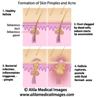
Skin acne formation, labeled diagram.
Anatomy of human skin showing process of formation of skin acne and pimples.
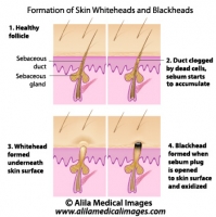
Skin blackheads illustration
Formation of skin blackhead and whitehead, labeled diagram.
Cellulite versus smooth skin, labeled diagram.
Illustration of normal skin anatomy (epidermis, dermis and subcutaneous fat) and changes in skin with cellulite : accumulation of fat cells push the fibrous septae up resulting in skin dimpling - bumps. Also, collagen fibers are less in number and less organised.
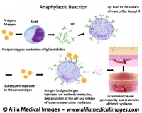
Mechanism of allergy (drug,hay fever, asthma...)
Anaphylactic reaction mechanism: antigen bridges the gap between two antibody molecules on mast cell or basophil, triggering massive release of histamine and other mediators.
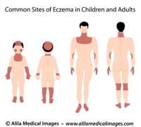
Sites of eczema on body, medical drawing.
Common sites of eczema in children and adults, medical illustration unlabeled.
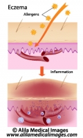
Eczema, allergic reaction, labeled diagram.
Allergens induce inflammation response of skin causing rash, redness and swelling.
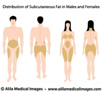
Subcutaneous fat distribution diagram.
Subcutaneous fat distribution in men and women, medical illustration.
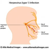
Herpes simplex virus infection, labeled diagram.
Herpes simplex virus (HSV-1) latent (in trigeminal nerve ganglion) and active infection (cold sores), labeled drawing.
Human skin anatomy, labeled.
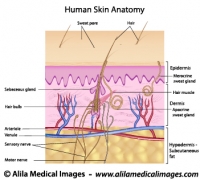
Structure of human skin, epidermis, dermis and hypodermis layers with hair follicle, sebaceous glands, nerve endings and blood supply.
Skin aging, medical illustration.
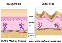
Older skin with wrinkles, age spots, dermis and hypodermis atrophy, reduced and disorganized collagen fibers.
Human skin anatomy, unlabeled.
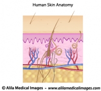
Structure of human skin, epidermis, dermis and hypodermis layers with hair follicle, sebaceous glands, nerve endings and blood supply.
Skin cancer staging, medical drawing.
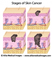
Melanoma of skin, staging based on layers affected (epidermis, dermis, hypodermis) and presence of metastasis, unlabeled.
Stages of pressure sores, labeled diagram.
Anatomy of skin layers and soft tissue showing different degrees of bed sores. Labeled illustration.
Degrees of skin burns, medical illustration.
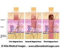
First degree of burn affect mostly the epidermis, second degree affects the dermis and third goes deeper into the hypodermis and below.
Contact dermatitis, medical drawing.
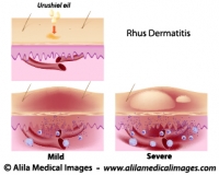
Urushiol oil induced contact (Rhus) dermatitis, illustration of the allergic reaction, unlabeled.


Email this to someoneShare on FacebookTweet about this on TwitterShare on Google+Share on LinkedIn