Tag Archives: pictures

Glaucoma

Below is a narrated animation about glaucoma development and types of glaucoma. Click here to license this video and/or other eye and vision related videos on Alila Medical Media website.

Glaucoma is a group of eye diseases in which the optic nerve is damaged leading to irreversible loss of vision. In most cases, this damage is due to an increased pressure within the eye – elevated intraocular pressure.

How glaucoma develops


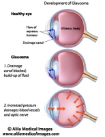
The eye produces a fluid called aqueous humor which is secreted by the ciliary body into the posterior chamber, a space between the iris and the lens. It then flows through the pupil into the anterior chamber between the iris and the cornea. From here, it is drained through a sponge-like structure located at the base of the iris called the trabecular meshwork and leaves the eye. In a healthy eye, the rate of secretion balances the rate of drainage.

In people with glaucoma, this drainage canal is partially or completely blocked. Fluid builds up in the chambers and this increases pressure within the eye. The pressure drives the lens back and presses on the vitreous body which in turn compresses and damages the blood vessels and nerve fibers running at the back of the eye. These damaged fibers result in patches of vision loss and if left untreated may lead to total blindness.

For eye anatomy basics click here.

Click here to see an animation of glaucoma progression on Alila Medical Media website where the video is also available for licensing.
Stages of glaucoma, a common eye disease
Fig. 1 : Development of glaucoma. Click on image to see a larger version on Alila Medical Media website where the image is also available for licensing.


Open-Angle Glaucoma vs. Angle-Closure Glaucoma


These are the two main types of glaucoma. The “angle” here refers to the corner between the cornea and the iris where the trabecular meshwork is located.

Primary Open-Angle or Chronic Open-Angle Glaucoma is the most common form of glaucoma accounting for about 90% of cases. This is caused by partial blockage of the drainage canal. The angle is “open”, meaning the entrance to the drain is clear, but the flow of aqueous humor is somewhat slow. The pressure builds up gradually in the eye over a long period of time. There is no pain and visual loss appears gradually, starting from peripheral vision, and may go on unnoticed until the central vision is affected. Progression of glaucoma can be stopped with medical treatments, but part of vision that is already lost can not be restored. This is why it’s very important to detect signs of glaucoma early with regular eye exams.

Closed-angle or Acute angle-closure glaucoma (AACG) is less common. This type of glaucoma is caused by a sudden and complete blockage of aqueous humor drainage. The pressure within the eye rises rapidly and may lead to total vision loss quickly. This is a medical emergency and requires immediate attention. Symptoms to watch out for: sudden severe pain inside and around the eye, redness, blurry vision, seeing halos around a light, some people may also feel headache, nausea.

Certain anatomical features of the eye make it easier for AACG to happen. These include: narrow drainage angle, shallow anterior chamber, thin and droopy iris, lens sitting too much forward. These features are often inherited and so AACG incidents are likely to run in the family.

Glaucoma closed angle vs open angle

Fig. 2 : Open angle vs closed angle glaucoma. Click on image to see a larger version on Alila Medical Media website where the image is also available for licensing.


Typically, this is what happens in AACG : the pupil is dilated (e.g. when looking in the dark) and the lens is stick to the back of the iris. This prevents the aqueous humor from flowing through the pupil into the anterior chamber (primary block). As the fluid accumulates in the posterior chamber it presses on the iris causing it to bulge outward and block the drainage angle (secondary block).

Other types of Glaucoma

Normal pressure glaucoma – Some people can get glaucoma (vision loss due to damaged optic nerve) without elevated intraocular pressure. This may be due to poor blood supply (e.g. damaged blood vessels in diseases such as diabetes) to the nerve fibers.

Secondary glaucoma – glaucoma develops as a result of trauma after eye injuries.

Congenital glaucoma – glaucoma that is present at birth.

Treatments

Progression of glaucoma can be halted or slowed down with medical treatments, but part of vision that is already lost can not be recovered. This is why it’s very important to detect signs of glaucoma early with regular eye exams.

Eye drops that lower intraocular pressure and/or reduce fluid production.

Laser treatments : Laser is used to burn part of the trabecular meshwork to improve fluid flow – laser trabeculoplasty. It can also be used to remove part of the ciliary body to reduce fluid secretion. For acute glaucoma, small holes can be made in the iris to relieve the primary block – laser iridotomy.

Eye surgeries: a procedure called trabeculectomy is used to create a channel –  an alternative route – for aqueous fluid drainage. For acute glaucoma a procedure called  iridectomy may be performed to drill a hole in the iris. Canaloplasty is a newer, less invasive surgical procedure performed for treatment of open angle glaucoma. This procedure involves enlargement of the eye’s natural drainage canal.

In people with AACG, laser and surgical treatments may be performed for the other, still healthy eye as well to prevent future development of glaucoma.

Email this to someoneShare on FacebookTweet about this on TwitterShare on Google+Share on LinkedIn

Reverse Total Shoulder Replacement

The videos on this page can be downloaded upon purchase of a license on Alila medical Media website. Click here!

Reverse Total Shoulder Replacement or Delta Shoulder Replacement is a modification of total shoulder replacement procedure. The conventional procedure has a metal ball on the humerus (replacing the natural humeral head) and a plastic component on the glenoid. The reverse procedure has the reverse positions of the prostheses : the metal ball is now fixed on the glenoid socket and the plastic cup is fixed in place of the humeral head (Fig. 1). This configuration is indicated for people with rotator cuff  arthropathy.
Conventional and reverse total shoulder replacement
Fig. 1: Configuration of conventional versus reverse total shoulder replacement. Note the location of the metal head and plastic component in each case. Click on image to see a larger version on Alila Medical Media website where the image is also available for licensing.


Why a conventional total shoulder will not work with rotator cuff  arthropathy?

Rotator cuff arthropathy or cuff tear arthropathy (CTA) is shoulder arthritis in a setting of rotator cuff tears. Rotator cuff muscles hold the ball and the socket together and prevent the humeral head from moving out of the socket when the deltoid muscle raises the arm. In people with torn rotator cuff, the head of the humerus moves upwards out of the socket resulting in shoulder instability. Conventional total shoulder preserving the natural configuration of the shoulder joint will encounter the same problem (Fig. 2, left panel).

Shoulder replacement options for cuff tear.


Fig. 2: Conventional versus reverse total shoulder replacement with torn rotator cuff. The upward force displaces the humeral head in conventional configuration but stabilizes the joint in reverse configuration. Click on image to see a larger version on Alila Medical Media website where the image is also available for licensing.


Why a reverse total shoulder would work?

In reverse total shoulder procedure, the arthritic humeral head is replaced with a plastic cup and the metal ball is fixed onto the glenoid surface. With this configuration, the contraction of the deltoid will move the arm up and compress the socket to the ball at the same time (Fig. 2, right panel). The ball and the socket will fit more snugly together providing stability.

                                                                                                            >  See all Orthopedic topics

Email this to someoneShare on FacebookTweet about this on TwitterShare on Google+Share on LinkedIn

Common knee injuries and surgical repair, part 1

Knee anatomy

The knee has three bones : the femur (the thigh bone), the tibia (the shinbone) and the patella (the kneecap). The femur and the tibia form a hinge joint. The joint is enclosed by the joint capsule at the back and on the sides, and is covered by the patella and patellar ligament in front. The knee joint is stabilized mainly by the tendons of quadriceps femoris muscle in front and semimembranosus muscle (one of the hamstrings) on the back. Strengthening these two muscles  therefore helps to reduce the risk of knee injuries.

Knee joint labeled drawing.

Fig.1 : Midsagittal section of the knee joint. Click on image to see a larger version on Alila Medical Media website where the image is also available for licensing.


Apart from the regular articular cartilage that cover the end surfaces of the three bones there are two additional pads of cartilage that are unique to the knee joint : medial meniscus and lateral meniscus (Fig. 2 and Fig. 3). The menisci act as shock absorbers to cushion the joint.

Two pairs of ligaments help to stabilize the knee : collateral ligaments run along two sides of the knee (Fig. 2), and cruciate ligaments which connect the femur and tibia in the center of the joint and cross each other in the from of an X (hence the names) : anterior cruciate ligament (ACL) and posterior cruciate ligament (PCL) (Fig. 2 and Fig. 3).

Knee joint anatomy

Fig. 2 : Front view of the right knee (the kneecap is removed in this picture to show structures behind).  Click on image to see a larger version on Alila Medical Media website where the image is also available for licensing.


Knee meniscus labeled diagram.
Fig. 3 : The right knee viewed from top (femur removed to show structures underneath). Click on image to see a larger version on Alila Medical Media website where the image is also available for licensing.


Meniscus tear and repair

Commonly referred to as torn cartilage, torn meniscus is a common sport injury. Mild injuries maybe treated with rest, ice, compression and elevation (the RICE approach). Larger tears may require surgery. The goal of surgery is to remove the damaged tissue which is the source of irritation, pain and possibly inflammation, and attempt to induce healing. Treatments vary depending on the location of the tear. If the tear is located on the outer border of the meniscus (the red zone, see Fig. 3) where there is a good blood supply, the damaged loose tissue will be removed and sutures will be used to tight the cartilage together to facilitate self healing. If the tear is located on the inner part (the white zone, see Fig. 3) where the blood supply is poor and hence healing is unlikely, the damaged part is simply removed (partial meniscectomy) (Fig. 4).

Minimal invasive arthroscopic surgery is commonly used for meniscus repair. In case of large tears, open surgery may be required.

Click here to see an animation of arthroscopic meniscus repair on Alila Medical Media website where the video is also available for licensing.

Meniscus tear and surgery treatment
Fig. 4 : Treatment of meniscus tear depends on its location. Click on image to see a larger version on Alila Medical Media website where the image is also available for licensing.


                                                                                                              >  See all Orthopedic topics


Email this to someoneShare on FacebookTweet about this on TwitterShare on Google+Share on LinkedIn

Common shoulder injuries and surgical repair (part 1)

Anatomy


There are three bones in the shoulder: the humerus (the bone of the upper arm), the scapula (shoulder blade) and the clavicle (collarbone). Articulations between these bones make up the shoulder joints. The main joint, commonly referred to as “the shoulder joint”, is the joint between the head of the humerus and glenoid cavity of the scapula and is called the humeroscapular or glenohumeral joint. The second joint of the shoulder is formed by the articulation between the clavicle and the acromion (extension of the scapula that forms the top of the shoulder) and is called acromioclavicular joint or AC joint. The two joints are stabilized by associated muscles and ligaments.

Shoulder anatomy
Fig.1: Main components of the shoulder joint. Click on image to see a larger version on  Alila Medical Media website where the image is also available for licensing.


Shoulder dislocation

Shoulder dislocation occurs when the humeral head slips out of the pocket  made by glenoid cavity of the scapula (Fig. 2). This usually happens as a result of trauma (fall, sport injury,..). Dislocation can be anterior where the humerus slips to the front or posterior where it dislocates behind the normal position. Anterior dislocation is more common.

Shoulder dislocation
Fig.2 : Types of shoulder dislocation. Click on image to see a larger version on Alila Medical Media website where the image is also available for licensing.


Below is a narrated animation about shoulder dislocation, bankart lesion and repair. Click here to license this video and/or other orthopaedic videos on Alila Medical Media website.

Bankart lesion and shoulder instability

The glenoid cavity has a ring of fibrocartilage tissue called the labrum around it. The labrum makes the cavity deeper and helps to keep the humeral head in place (Fig.3). During anterior shoulder dislocation, the head of the humerus may be pressed against and damages the anterior portion of the labrum. This type of labral tear is called bankart. Damaged labrum makes it easier for the humeral head to slip out of place again. This vicious cycle leads to repeated shoulder dislocation and severely damaged labrum. The condition is called shoulder instability as it feels like slipping out anytime. Treatment includes physical therapy and, in some cases, surgery for bankart repair (see below).

Click here to see an animation of bankart lesion and arthroscopic repair  on Alila Medical Media website where the video is also available for licensing.

SLAP and bankart lesions
Fig. 3: Anatomy of the shoulder joint with the humerus slightly abducted to show the glenoid cavity and labrum. Types of labral lesions are shown on the right. Click on image to see a larger version on Alila Medical Media website where the image is also available for licensing.


Bankart repair

During surgery the damaged cartilage is removed, area is cleaned, small holes are then drilled into the bone of glenoid fossa to hold small suture anchors with threads. The threads are attached to the labrum and pulled tightly to hold the labrum to the glenoid (Fig.4). Over time, the labrum will reattach to the glenoid naturally. Physical therapy will be needed to regain the shoulder range of motions and strength.

Shoulder stabilization surgery
Fig.4: Steps of bankart repair surgery. See text for details. Click on image to see a larger version on Alila Medical Media website where the image is also available for licensing.


Bankart repair can be done with arthroscopic or open surgery. While arthroscopy is minimal invasive, open surgery maybe recommended for larger tears. In arthroscopic surgery only two or three small incisions are made, an arthroscope is inserted through one of the incision. Arthroscope is an instrument equipped with light and camera which transmits image of the joint to a computer screen. Other small tools are inserted  to carry out the repair (Fig.5).

Click here to see an animation of arthroscopic bankart repair.
Shoulder arthroscopy
Fig. 5 : Arthroscopic surgery for bankart repair. Click on image to see a larger version on Alila Medical Media website where the image is also available for licensing.


                                                                                                              >  See all Orthopedic topics

Email this to someoneShare on FacebookTweet about this on TwitterShare on Google+Share on LinkedIn

Male Reproductive System Gallery

Download diagrams of male organs anatomy and diseases, formation and structure of sperms.

Please note: Free downloads are intended to facilitate healthcare education for people in need in low income countries and can be used for educational purposes only. If you can afford it or if you plan to use the images for commercial purposes, please consider buying instead. You can find a larger image collection at higher resolution for sale at affordable prices on Alila Medical Media website.

To download: right click on full size image – choose “Save image as” and save it into your computer. By downloading from this website you acknowledge that you agree to our Conditions of Use.
To purchase larger sizes of the image: (left) click on full size image.

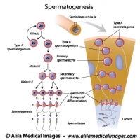
Spermatogenesis process, labeled diagram.
Process of sperm production and meiosis in seminiferous tubule, labeled diagram.
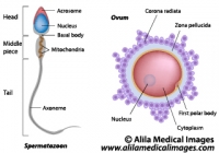
Ovum and spermatozoon structure, labeled diagram.
Egg and sperm cells anatomy, labeled drawing.
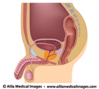
Male reproductive organs sagittal, unlabeled diagram.
Illustration of male reproductive system in median section
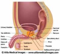
Male reproductive organs sagittal, labeled diagram.
Illustration of male reproductive system in median section
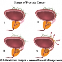
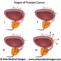
Prostate cancer staging, unlabeled diagram.
Illustration of staging of prostate gland cancer, from small tumor size to metastasis.
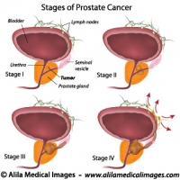
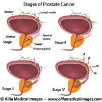
Prostate cancer staging, labeled diagram.
Illustration of staging of prostate gland cancer, from small tumor size to metastasis.


Email this to someoneShare on FacebookTweet about this on TwitterShare on Google+Share on LinkedIn

Lymphatic System Gallery

Download illustrations of major groups of lymph nodes, lymphatic organs and vessels, their anatomy, function, disorders and involvement in cancer metastasis.

Please note: Free downloads are intended to facilitate healthcare education for people in need in low income countries and can be used for educational purposes only. If you can afford it or if you plan to use the images for commercial purposes, please consider buying instead. You can find a larger image collection at higher resolution for sale at affordable prices on Alila Medical Media website.

To download: right click on full size image – choose “Save image as” and save it into your computer. By downloading from this website you acknowledge that you agree to our Conditions of Use.
To purchase larger sizes of the image: (left) click on full size image.

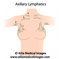
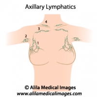
Axillary lymph nodes, medical drawing.
Axillary lymph nodes diagram, simple version. 1-3 = different groups of axillary nodes, 4= supraclavicular lymph nodes. Unlabeled diagram.
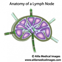
Lymph node structure, unlabeled diagram.
Diagram illustrating structure of a lymph node with lymph flow, unlabeled.
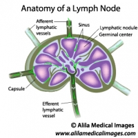
Lymph node structure, labeled diagram.
Diagram illustrating structure of a lymph node with lymph flow, labeled.
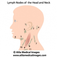
Lymph nodes of head and neck, medical drawing.
Lymph nodes of the head and neck, unlabeled. 1= preauricular, 2= posterior auricular, 3= parotid, 4= occipital, 5= submandibular, 6= submental, 7-10 = cervical, 11= supraclavicular.
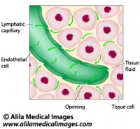
Lymphatic fluid drainage, labeled diagram.
Uptake of tissue fluid by lymphatic capillary, labeled drawing.
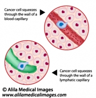
Metastatic cancer, medical drawing.
Cancer cell squeezes through the endothelium of blood and lymph vessel, illustration unlabeled.
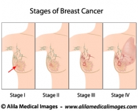
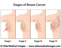
Breast cancer staging, diagram.
Stages of Breast Cancer, from small tumor to metastasis, medical illustration, unlabeled.
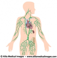
Lymphatic system, unlabeled diagram.
Human lymphatic system with all organs, major trunks, ducts and groups of lymph nodes illustrated, unlabeled illustration.
Lymphatic system, labeled diagram.
Human lymphatic system with all organs, major trunks, ducts and groups of lymph nodes illustrated, labeled illustration.


Email this to someoneShare on FacebookTweet about this on TwitterShare on Google+Share on LinkedIn

Female Reproductive System Gallery

Download cliparts illustrating anatomy of reproduction organs, oogenesis and sexual cycle, hormonal control, women’s diseases and conditions.

Please note: Free downloads are intended to facilitate healthcare education for people in need in low income countries and can be used for educational purposes only. If you can afford it or if you plan to use the images for commercial purposes, please consider buying instead. You can find a larger image collection at higher resolution for sale at affordable prices on Alila Medical Media website.

To download: right click on full size image – choose “Save image as” and save it into your computer. By downloading from this website you acknowledge that you agree to our Conditions of Use.
To purchase larger sizes of the image: (left) click on full size image.

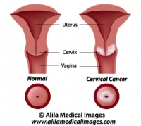
Cervical cancer, labeled diagram.
Diagram showing cancer of the cervix, labeled.
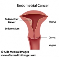
Uterine cancer, labeled diagram.
Illustration of tumor of the endometrium of female uterus, labeled.
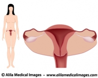
Women's system simple version, unlabeled.
Diagram showing female body with anatomy of reproductive system, anterior view, simple unlabeled version.
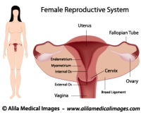
Women's system simple version, labeled.
Diagram showing female body with anatomy of reproductive system, anterior view, simple labeled version.
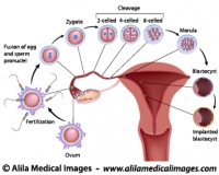
From fertilization to implantation, labeled diagram.
Diagram showing early human embryo development events, from fertilization to implantation of blastocyst, labeled.
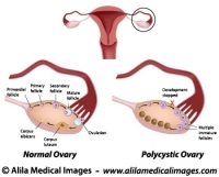
Polycystic and normal ovary, labeled diagram.
Normal ovarian cycle and Polycystic ovary syndrome illustration, labeled.
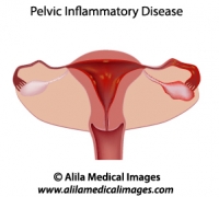
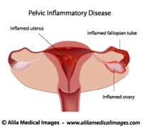
Pelvic inflammatory disease (PID)
Pelvic inflammatory disease (PID) diagram, unlabeled
Pelvic inflammatory disease (PID) diagram
Pelvic inflammatory disease (PID) diagram, labeled
Axillary lymph nodes, medical drawing.
Axillary lymph nodes diagram, simple version. 1-3 = different groups of axillary nodes, 4= supraclavicular lymph nodes. Unlabeled diagram.
Breast cancer staging, diagram.
Stages of Breast Cancer, from small tumor to metastasis, medical illustration, unlabeled.
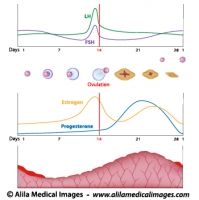
Menstrual cycle and ovulation, labeled diagram.
Normal menstrual cycle and the hormonal control of ovulation, labeled illustration.
Women's reproductive system, unlabeled diargam.
Diagram showing organs of female reproductive tract in median section.
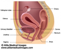
Women's reproductive system, labeled diargam.
Diagram showing organs of female reproductive tract in median section.
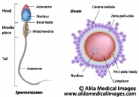
Ovum and spermatozoon structure, labeled diagram.
Egg and sperm cells anatomy, labeled drawing.


Email this to someoneShare on FacebookTweet about this on TwitterShare on Google+Share on LinkedIn

Oral Cavity Gallery

Download illustrations of permanent and deciduous dentition, tooth anatomy, periodontal diseases, teeth replacement therapy, mouth anatomy and diseases.

Please note: Free downloads are intended to facilitate healthcare education for people in need in low income countries and can be used for educational purposes only. If you can afford it or if you plan to use the images for commercial purposes, please consider buying instead. You can find a larger image collection at higher resolution for sale at affordable prices on Alila Medical Media website.

To download: right click on full size image – choose “Save image as” and save it into your computer. By downloading from this website you acknowledge that you agree to our Conditions of Use.
To purchase larger sizes of the image: (left) click on full size image.

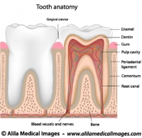
Tooth anatomy, unlabeled diagram.
Tooth anatomy from outside and in cross section, unlabeled drawing.
Tooth anatomy, labeled diagram.
Tooth anatomy from outside and in cross section, labeled drawing.
Dental checkup icon
Dental checkup icon
Magnifying glass on a molar tooth, illustration.
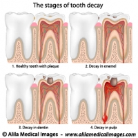
Plaque and tooth decay, medical drawing.
Tooth decay stages diagram.
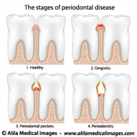
Periodontal Disease, labeled diagram.
Gum disease stages, from gingivitis to periodontitis.
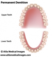
Adult dentition, medical drawing.
Permanent teeth, adult dentition, upper and lower jaws, unlabeled.
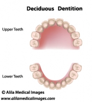
Deciduous dentition, medical drawing.
Deciduous dentition (baby or milk teeth), upper and lower jaws, medical illustration unlabeled..
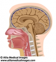
Head anatomy median section, medical drawing.
Median section of human head diagram with details of brain, sinuses, tonsils, oral and nasal cavity, pharynx and larynx, unlabeled.
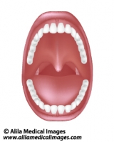
Mouth and throat anatomy, medical drawing.
Diagram of human oral cavity with denture, tonsils, unlabeled.


Email this to someoneShare on FacebookTweet about this on TwitterShare on Google+Share on LinkedIn

Urinary System Gallery

Download anatomical illustrations of kidneys, urine formation process, urinary tract diseases and conditions.

Please note: Free downloads are intended to facilitate healthcare education for people in need in low income countries and can be used for educational purposes only. If you can afford it or if you plan to use the images for commercial purposes, please consider buying instead. You can find a larger image collection at higher resolution for sale at affordable prices on Alila Medical Media website.

To download: right click on full size image – choose “Save image as” and save it into your computer. By downloading from this website you acknowledge that you agree to our Conditions of Use.

To purchase larger sizes of the image: (left) click on full size image.

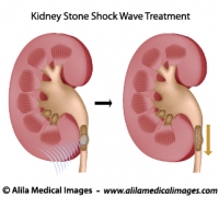
Treatment of kidney stones, medical drawing.
Ultrasound shock wave therapy for kidney stones, unlabeled.
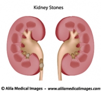
Kidney stones, medical drawing.
Diagram of healthy kidney anatomy and kidney with stones blocking urine passage, unlabeled.
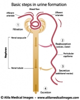
Formation of urine, labeled diagram.
Basic steps in urine formation in a nephron of kidney.
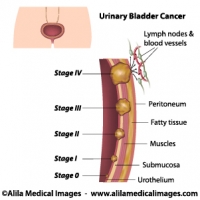
Urinary bladder cancer stages, medical drawing.
Staging of urinary bladder cancer diagram, unlabeled
Urinary bladder cancer stages, labeled.
Staging of urinary bladder cancer diagram, labeled
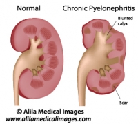
Chronic pyelonephritis, labeled diagram.
Chronic pyelonephritis kidney disease with tissue scarring and blunted calyx, medical illustration.
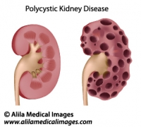
Polycystic kidney disease, medical drawing.
Diagram of enlarged kidney with multiple cysts compared to normal anatomy, unlabeled.
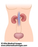
Urinary organs in female, medical illustration.
Female urinary system diagram showing kidneys, ureters and bladder, simple version unlabeled.
Prostate cancer staging, unlabeled diagram.
Illustration of staging of prostate gland cancer, from small tumor size to metastasis.
Prostate cancer staging, labeled diagram.
Illustration of staging of prostate gland cancer, from small tumor size to metastasis.


Email this to someoneShare on FacebookTweet about this on TwitterShare on Google+Share on LinkedIn

Eyes, Vision Gallery

Download diagrams of human eye anatomy, tear film and lacrimal gland, muscles of the orbit and associated nerves, defects and diseases, laser surgery treatment.

Please note: Free downloads are intended to facilitate healthcare education for people in need in low income countries and can be used for educational purposes only. If you can afford it or if you plan to use the images for commercial purposes, please consider buying instead. You can find a larger image collection at higher resolution for sale at affordable prices on Alila Medical Media website.

To download: right click on full size image – choose “Save image as” and save it into your computer. By downloading from this website you acknowledge that you agree to our Conditions of Use.
To purchase larger sizes of the image: (left) click on full size image.

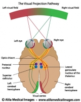
Visual pathways , labeled diagram.
Diagram of visual projection pathway from eyes to brain (visual cortex), labeled.
Visual pathways , unlabeled diagram.
Diagram of visual projection pathway from eyes to brain (visual cortex), unlabeled.
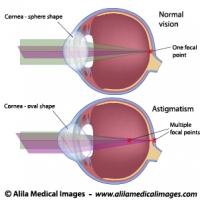
Astigmatism, labeled diagram.
The oval shape of the cornea of the eye in people with astigmatism creates multiple focal points.
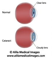
Cataract, labeled diagram.
Illustration of Eye disease - Cataract, clouding of lens.
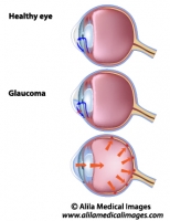
Glaucoma, unlabeled diagram.
Illustration of stages of development of glaucoma, a common eye disease.
Glaucoma, labeled diagram.
Illustration of stages of development of glaucoma, a common eye disease.
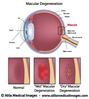
Macular degeneration, labeled diagram.
Diagram showing eye common disease: macular degeneration (dry and wet), labeled.
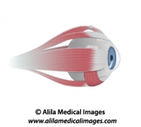
Muscles of the eye, unlabeled diagram.
Diagram showing extrinsic muscles controlling movement of the eye, unlabeled.
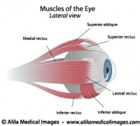
Muscles of the eye, labeled diagram.
Diagram showing extrinsic muscles controlling movement of the eye, labeled.
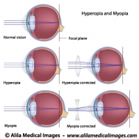
Common eye defects diagrams
Myopia, hyperopia and correction with lenses, labeled illustrations.
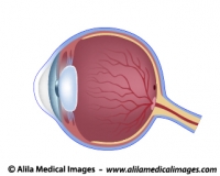
Eye anatomy unlabeled diagram.
Cross section of human eye illustration, unlabeled.
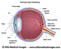
Eye anatomy labeled diagram.
Cross section of human eye illustration, labeled.
Eye adaptation to near vision, illustration.
The near response of the eye , lens accommodation, labeled diagram.
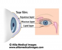
Dry eye syndrome, labeled diagram
Tears composition and the glands involved in tears production, labeled.


Email this to someoneShare on FacebookTweet about this on TwitterShare on Google+Share on LinkedIn